|

|
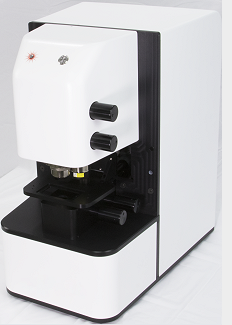
ImageLab for Spero is a one-of-a-kind image processing tool for the analysis of QCL-IR hyperspectral image cubes obtained by Daylight's Spero Chemical Imaging Microscope. The many on-board tools, based on multivariate statistics, enable the user to analyze and classify the hyperspectral images. This ultimately enables users to merge the spectral data with high-resolution microscope photos.
Furthermore, ImageLab for Spero provides a user-friendly programming language that can be used to automate analyses of the hyperspectral image data. To learn more about the power of ImageLab, please visit our collection of how-to videos and webinars.
|
 |
Unique Features
- One-of-a-kind image processing tool specifically tailored to fit the Spero instrument
- Simple and flexible data import
- A large built-in library of statistical methods enabling advanced chemometric analysis of hyperspectral images:
- Principal Component Analysis (PCA)
- Cluster Analysis (dendrograms, k-means and fuzzy c-means)
- Correlation/Similarity Maps (SIMAP)
- PLS Discriminant Analysis (PLS/DA)
- Random Decision Forest (RDF)
- Vertex Component Analysis (VCA, endmember detection)
- Maximum Noise Fraction Transform (MNF)
- ImageLab for Spero is fully programmable using the built-in ILabPascal compiler to create user-specific analyses for routine tasks.
- A flexible and easy-to-learn user interface that flattens the learning curve and guarantees immediate success in the analysis and visualization of information.
- Spectral preprocessing: spectral scaling, baseline correction, CO2 peak removal, 1st and 2nd derivative, smoothing, and more.
- Spatial preprocessing: lowpass and highpass filtering, Sobel and Laplace filters, Hough circle transform, resampling, and more.
- Spectral descriptors allow for fine tuning the selectivity and sensitivity of chemometric models.
- A flexible annotation tool allows to annotate both spectra and images.
- Automatic particle detection (applied both to raw spectra and classified images).
- Up to eight raw or processed images and up to four image cubes from different sources (e.g. QCL-IR + Raman + visible) can be stacked or merged for multi-spectral data visualization or in highlighting a region of interest with false-color tags.
|
|
The power of ImageLab for Spero can also be seen from the high number of scientific publications which have been published so far. Following are a few papers specifically created with the help of ImageLab for Spero:
|
|
| L. Shi, X. Liu, L. Shi, H.T. Stinson, J. Rowlette, L.J. Kahl, C.R. Evans, C. Zheng, L.E.P. Dietrich, W. Min |
Mid-infrared metabolic imaging with vibrational probes.
Nature Methods 17, 844851 (2020). DOI: 10.1038/s41592-020-0883-z |
| B. Bird, J. Rowlette |
High definition infrared chemical imaging of colorectal tissue using a Spero QCL microscope.
Analyst, Jan 2017, DOI: 10.1039/c6an01916a.
|
| B. Bird, J. Rowlette |
A protocol for rapid, label-free histochemical imaging of fibrotic liver.
Analyst, Nov 2016, DOI: 10.1039/c6an02080a.
|
| B. Bird |
Large-area infrared chemical imaging of colorectal tissue using a Spero microscope.
Application Note 201602, DayLight Solutions Inc.
|
| B. Mohar |
Real-time mid-IR chemical imaging of dynamic processes: Proton-Deuteron exchange within a microfluidic system using the Spero(TM) QCL based microscope.
Application Note: 201501, Daylight Solutions Inc.
|
|
|