
Data Repository
The following collection of datasets has been prepared for Epina ImageLab, the files can be loaded directly into Epina ImageLab(1).
| Imaging Method(s) | Image Size | Spectral Size | Download | |
| DS001 Coffee beans mixed with stones |
near infrared | 319x359 | 156 | coffee_mix1022.zip [69 MB] 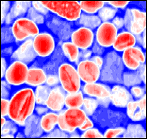 |
| This remarkable dataset contains the hyperspectral image of a mixture of coffee beans and rocks. The spectra exhibit lots of spikes and saturated regions which form an excellent test case for algorithms detecting and correcting artifacts in hyperspectral images. Many thanks to Viktoria Parrag for providing the data; Reference: Parrag, V; Felföldi, J; Firtha, F (2014): Roasted and green coffee beans with stones: a hyperspectral training data set for multivariate image analysis. CORVINUS University of Budapest, Department of Physics and Control, Faculty of Food Science. |
||||
| DS002 Concrete |
mass spectrometry | 40x50 | 8 | concrete.zip [1.5 MB]  |
| Concrete sample taken from the wall of an road tunnel. MS image acquired by laser ablation in combination with ICP/MS; eight specific isotopes have been measured: 13C, 25Mg, 27Al, 29Si, 35Cl, 42Ca, 49Ti, and 57Fe. Data courtesy Andreas Limbeck, Vienna Univ. of Technology. |
||||
| DS003 Dust |
EDX (energy dispersive xray) + Raman | 101x101 | 12 (EDX) + 1024 (Raman) | dust.zip [61 MB] 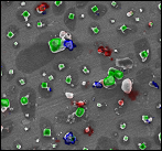 |
| A multisensor dataset which combines energy dispersice xray (EDX) and Raman spectra of a sample of particulate matter obtained in the salt lake district of Western Australia. Data courtesy Johannes Ofner, Vienna University of Technology. Reference: H. Lohninger, J. Ofner: Multisensor hyperspectral imaging as a versatile tool for image-based chemical structure determination. Spectroscopy Europe Vol. 26 No. 5 (2014), 6-10´. |
||||
| DS004 Gulf of Mexico Wetlands |
visible & near infrared | 320x401 | 360 | spectir_wetlands_p2.zip [64 MB]  |
| This data sample is taken from SpecTIRs extensive hyperspectral baseline environmental dataset covering multiple National Wildlife Reserves in the Gulf of Mexico during the period of May-June 2010. This specific sample is from the Lower Suwanee NWR, collected at a 2m spatial resolution and a nominal 5nm band spacing covering the spectral range of 395 nm to 2.0 µm. | ||||
| DS005 Atmospheric Particulate Matter |
EDX (energy dispersive xray, 12 elements + backscatter signal) Raman SEM |
101x101 | 13 (EDX, incl. backscatter) 1024 (Raman) |
atm_part_matter.zip [80 MB] 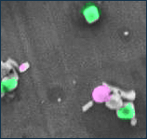 |
| This multisensor dataset contains data fused from three imaging methods (Raman, energy-dispersive x-ray, and scanning electron microscope). Data courtesy Johannes Ofner; Reference: J. Ofner, K.A. Kamilli, E. Eitenberger, G. Friedbacher, B. Lendl, A. Held, H. Lohninger: Chemometric analysis of multisensor hyperspectral images of precipitated atmospheric particulate matter. Analytical Chemistry 87 (2015) pp. 9413-9420 |
||||
| DS006 Keplercard |
THz |
48x48 | 768 | keplercard.zip [7.2 MB] 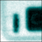 |
| An HSI of an electronic banking card showing the transponder chip and parts of the copper coil. The data contain both the THz spectra and the pulses for time domain spectroscopy. Data courtesy Harald Pühringer, Recendt GmbH, Linz, Austria. |
||||
| DS007 Paeonia |
mass spectrometry | 120x120 | 4 | paeonia_petal.zip [0.7 MB]  |
| Petal of a Paeonia which has been watered in water containing blue ink. The hyperspectral image has been obtained by LA-ICP-MS. Data courtesy Max Bonta of Vienna University of Technology, Inst. of Chemical Technologies and Analytics. |
||||
| DS008 Steel |
ToF-SIMS | 512x512 | 52 | sobol_unger_steeldata.zip [5.2 MB] 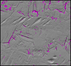 |
| The combination of ToF-SIMS and HRSEM allows the investigations of hydrogen embrittlement in duplex stainless steel microstructures. Data courtesy Wiley. Reference: O. Sobol, G. Holzlechner, M. Holzweber, H. Lohninger, T. Boellinghaus, W.E.S. Unger: "First Use of and Multivariate Analysis and Data Fusion of ToF-SIMS and HR-SEM image data for Studying Deuterium Assisted Degradation Processes in Duplex Steels", ECASIA 2015 Proceedings, to be published in Surface and Interface Analysis, Wiley 2016 |
||||
| DS009 Copper Sulfide Particles |
EDX + Raman + ToF-SIMS | 100x100 | 4 (EDX) + 294 (Raman) + 38 (ToF-SIMS) |
copper_sulfide_particles.zip [25.0 MB] 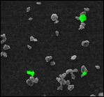 |
| The copper sufide in this sample contains a small amount of copper oxide, which lights up in green in the image at the right.
This dataset is part of a larger dataset collection (DS009...DS012) used in the following paper: Reference: J. Ofner et al.: Image-Based Chemical Structure Determination, Scientific Reports Vol. 7 (2017) 6832 |
||||
| DS010 Ceramics |
EDX + Raman + ToF-SIMS | 201x201 | 10 (EDX) + 228 (Raman) + 68 (ToF-SIMS) |
ceramic_composite.zip [96.3 MB] 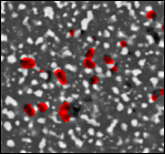 |
| This technical ceramic (ZrO2-reinforced mullite) shows tiny diamonds on top of its surface (red spots). The diamonds are a residue of the diamond polish paste used for the preparation of the sample. This dataset is part of a larger data collection (DS009...DS012) used in the following paper: Reference: J. Ofner et al.: Image-Based Chemical Structure Determination, Scientific Reports Vol. 7 (2017) 6832 |
||||
| DS011 Tumor Cells |
EDX + Raman + ToF-SIMS | 176x101 | 13 (EDX) + 335 (Raman) + 87 (ToF-SIMS) |
tumour_cells.zip [57.4 MB]  |
| Sample of two tumor cells. The image at the right shows a single cell with its nucleus encircled by the darkblue line which has been calculated from the Raman signal.
This dataset is part of a larger dataset collection (DS009...DS012) used in the following paper: Reference: J. Ofner et al.: Image-Based Chemical Structure Determination, Scientific Reports Vol. 7 (2017) 6832 |
||||
| DS012 Environmental Aerosol |
EDX + Raman + ToF-SIMS | 300x300 | 18 (EDX) + 682 (Raman) + 263 (ToF-SIMS) |
environmental_aerosol.zip [579.2 MB] 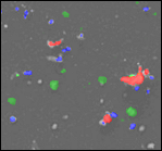 |
| Environmental sample showing sodium (green), calcium (red) and iron (blue) signals. This dataset is part of a larger dataset collection (DS009...DS012) used in the following paper: Reference: J. Ofner et al.: Image-Based Chemical Structure Determination, Scientific Reports Vol. 7 (2017) 6832 |
||||
| DS013 Concrete |
LIBS (Laser Induced Breakdown Spectroscopy) | 101x101 | 12288 | LIBS_concrete.zip [292.8 MB]  |
| Concrete sample showing full LIBS spectra between 200 and 1000 nm with 0.08 nm spectral resolution. Data courtesy A. Limbeck, TU Wien, Austria. |
||||
| DS014 Microplastic |
MID-IR | 295x276 | 1218 | microplastic.zip [403.7 MB] 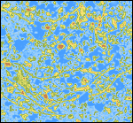 |
| A sample showing microparticles of different polymers. The dataset is demanding due to the strong Mie scattering effect. Data courtesy M. Löder & C. Laforsch, Univ. of Bayreuth, Germany. |
||||
| DS015 Euro Coin |
LIBS (Laser Induced Breakdown Spectroscopy) | 127x90 | 9610 | euro2_libs.zip [256 MB]  |
| A 2-Euro coin mapped by LIBS. The coin consists of nickel and brass, thus selecting either Ni, Zn or Cu lines exhibits the composition of the coin. Spectra are between 187 and 836 nm Data courtesy A. Limbeck, TU Wien, Austria. |
||||
| (1) | Please note: the datasets published on this page have been specially signed to allow full access to the data even with the evaluation copy of Epina ImageLab (required version: 1.01 or higher). Any change of the data or the corresponding meta information will render the digital signature invalid. |